X-rays hold a critical position in the realm of orthopedics, particularly in the diagnosis and treatment of bone and joint injuries. These imaging techniques have revolutionized the way orthopedic specialists understand and address musculoskeletal issues.
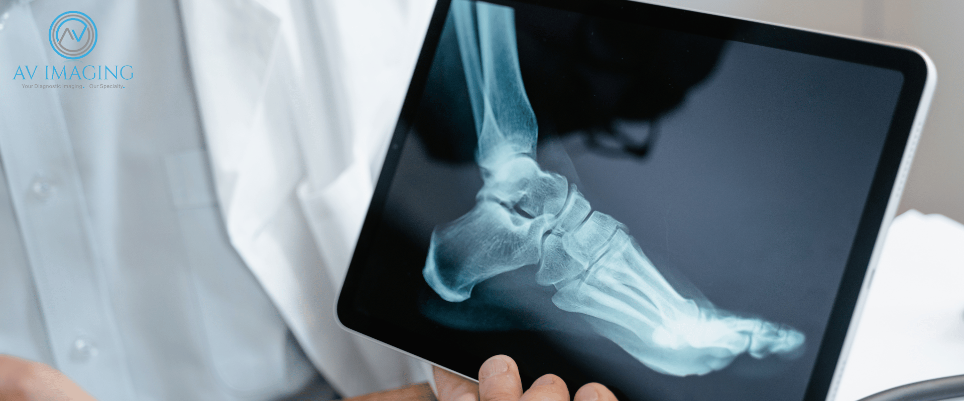
X-rays, a type of electromagnetic radiation, are instrumental in producing images of bones and joints. Orthopedic doctors use X-rays as a primary diagnostic tool to assess fractures, dislocations, arthritis, and other bone-related conditions. The images captured by X-rays enable physicians to visualize the structure of bones and detect abnormalities or injuries.
When a patient presents with a suspected bone or joint injury, X-rays are often the initial step in diagnosis. Patients are positioned accordingly, and X-ray machines emit radiation through the affected area. The resulting images showcase the affected bone's alignment, integrity, and any potential fractures or abnormalities.
X-rays are particularly effective in identifying fractures. They help orthopedic specialists determine the location, severity, and type of fracture, crucial information for devising an appropriate treatment plan. Whether it's a simple or complex fracture, X-rays provide detailed insights that aid in deciding the best course of action.
he information obtained from X-rays guides orthopedic surgeons in planning treatments. For instance, a simple fracture might require immobilization through casting, while complex fractures may necessitate surgical intervention. X-rays play a vital role in monitoring the progress of healing during follow-up visits.
While X-rays are invaluable, they do have limitations. They primarily depict bone structures and might not adequately visualize soft tissues like tendons or ligaments. However, advancements such as MRI or CT scans complement X-rays by offering detailed views of soft tissues when necessary.
Orthopedic professionals take precautions to limit radiation exposure during X-ray procedures. They use lead shielding and employ the lowest possible radiation dose to obtain clear images while ensuring patient safety.
Connect with us to learn more about how the AV Imaging team can help!